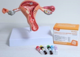
New autogenous tooth transplantation model—using 3D printing to assist with dental autotransplantation
Source: Taipei Medical University Hospital
Published on 2020-07-06
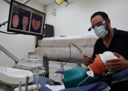
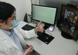
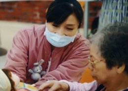
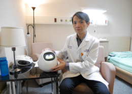
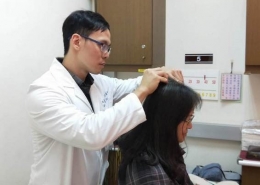
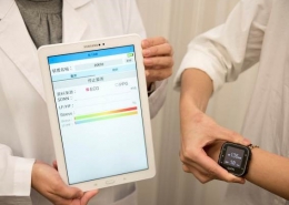
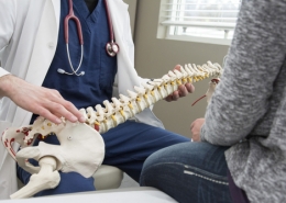
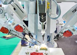
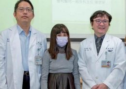
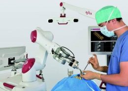
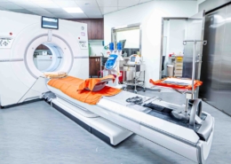
The Taipei Medical University Hospital is using 3D printing technology to help assist autogenous tooth transplantation.
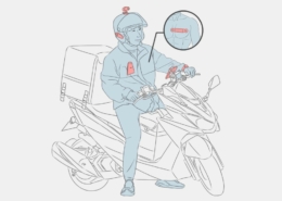
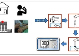
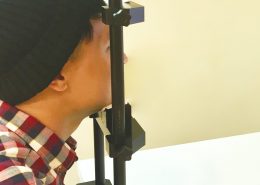
Prior to the transplantation surgery, a model of the tooth to be translated (called the donor tooth) can be made with 3D printing, and based on the shape of the model, the transplantation site on the alveolar bone can be trimmed to the correct size before the donor tooth is extracted and transplanted. This greatly reduces the time the donor tooth spends outside the oral cavity, saving precious time during the surgery and improves on the quality of the prognosis.
Did you know? |
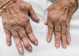
| Traditional autogenous tooth transplantation is to first fully remove the donor tooth, and trim the alveolar bone at the transplantation site based on the shape of the donor tooth, so that the shape of the bone is similar to the shape of the root of the tooth, before the donor tooth is transplanted and fixed in place. Due to the varying shapes of each tooth, the trimming can only be performed during the surgery, which invariably increases the amount of time the donor tooth spends exposed to the outside environment, which affects the success rate of dental autotransplantation. The longer the donor tooth spends outside the oral cavity, the easier it is to damage the nerves in the tooth. According to statistics, the golden window for transplantation is within 30 minutes of removal. The length of the transplantation surgery has significant impact on the prognosis of the surgery. |
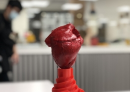
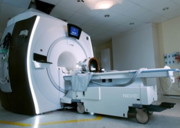
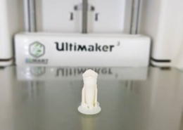
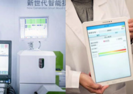
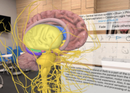
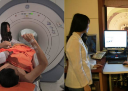
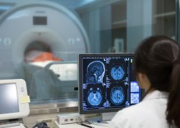
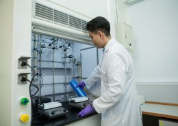
To reduce the waiting time of trimming of the alveolar bone after the donor tooth is extracted, TMU Hospital and TMU College of Oral Medicine collaborated on a R&D effort, where the newest 3D printing technology was used. Prior to the autotransplantation surgery, tomography is used to obtain the shape of the donor tooth, and 3D printing technology to generate a 1:1 ratio model of the donor tooth. During the surgery, the donor tooth is not extracted first; instead the model is used at the transplantation to trim the shape, until the site is the proper size before the donor tooth is extracted and transplanted. This greatly reduces the time the donor tooth spends outside the oral cavity. From the extraction of the donor tooth to the completion of the surgery, the process can be completed within three minutes.
The use of 3D printing technology can not only reduce the time donor tooth spend exposed to the outside environment, but can also reduce the wear and tear on the surface of the tooth root that occurs during the trimming and repeated testing of the match between the donor tooth and the transplantation site. This allows a greater degree of completeness of the tooth root surface, which is of great help to the stability of autogenous tooth transplantation, and reduces the likelihood of sequela.

Prior to the transplantation surgery, tomography is used to obtain the shape of the donor tooth, and 3D printing technology is used to generate a 1:1 donor tooth model
 https://oge.tmu.edu.tw/wp-content/uploads/2024/04/olympiad-poster-2.jpg
909
640
gps
/wp-content/uploads/2018/05/02-紅logo並置.png
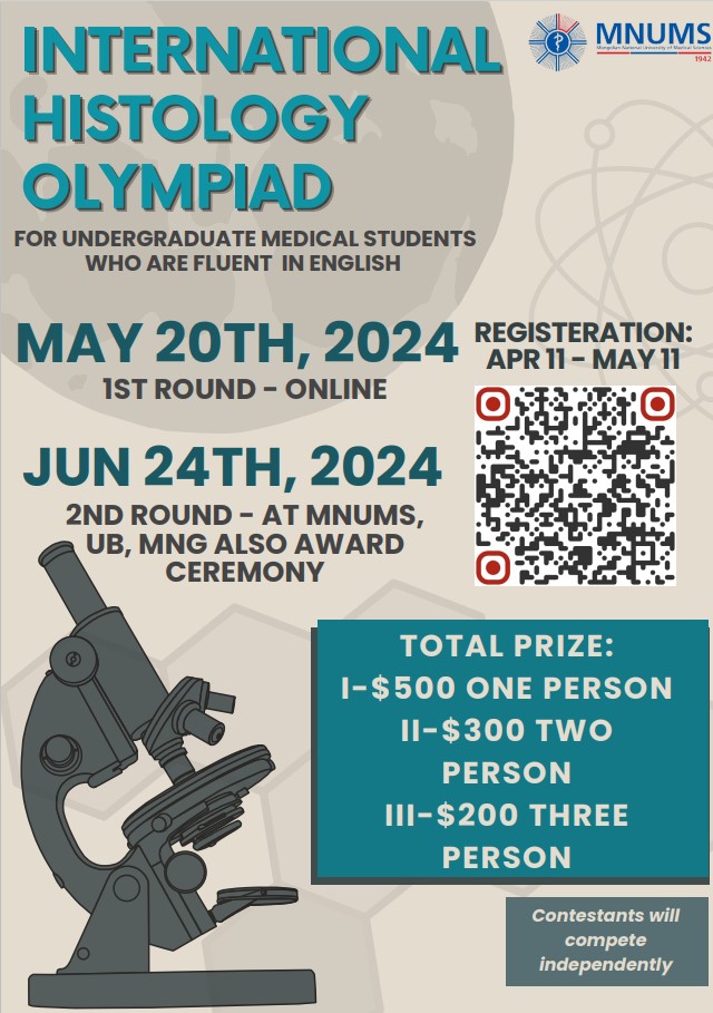
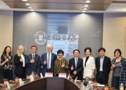
gps2024-04-19 16:36:372024-04-19 16:38:14International Histology Olympiad at the Mongolian National University of Medical Sciences (MNUMS)
https://oge.tmu.edu.tw/wp-content/uploads/2024/04/olympiad-poster-2.jpg
909
640
gps
/wp-content/uploads/2018/05/02-紅logo並置.png
gps2024-04-19 16:36:372024-04-19 16:38:14International Histology Olympiad at the Mongolian National University of Medical Sciences (MNUMS) https://oge.tmu.edu.tw/wp-content/uploads/2023/07/20230321台北醫學大學校園形象攝影-1-scaled.jpg
1000
1500
global.initiatives
/wp-content/uploads/2018/05/02-紅logo並置.png
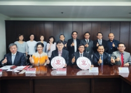
global.initiatives2024-04-19 16:08:552024-04-23 17:47:37[Open Application] Fall 2024 TMU admission for applicants seeking the MOFA Taiwan scholarship
https://oge.tmu.edu.tw/wp-content/uploads/2023/07/20230321台北醫學大學校園形象攝影-1-scaled.jpg
1000
1500
global.initiatives
/wp-content/uploads/2018/05/02-紅logo並置.png
global.initiatives2024-04-19 16:08:552024-04-23 17:47:37[Open Application] Fall 2024 TMU admission for applicants seeking the MOFA Taiwan scholarship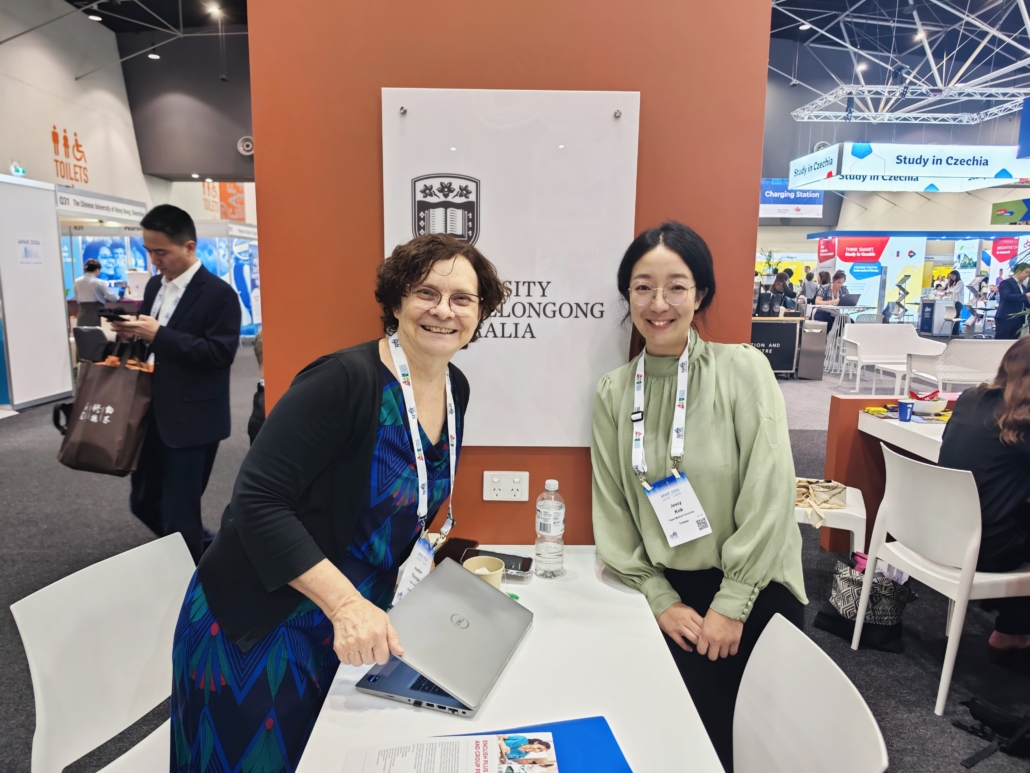 https://oge.tmu.edu.tw/wp-content/uploads/2024/04/1712888393174-scaled.jpg
1125
1500
gps
/wp-content/uploads/2018/05/02-紅logo並置.png
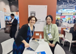
gps2024-04-15 16:31:512024-04-16 10:48:20Outward visit_2024 Asia Pacific Association of International Education (APAIE) conference and exhibition
https://oge.tmu.edu.tw/wp-content/uploads/2024/04/1712888393174-scaled.jpg
1125
1500
gps
/wp-content/uploads/2018/05/02-紅logo並置.png
gps2024-04-15 16:31:512024-04-16 10:48:20Outward visit_2024 Asia Pacific Association of International Education (APAIE) conference and exhibition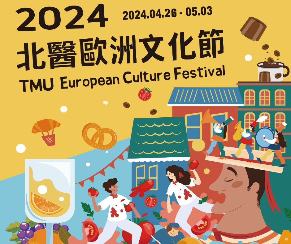 https://oge.tmu.edu.tw/wp-content/uploads/2024/04/for-OGEW.jpg
788
940
global.initiatives
/wp-content/uploads/2018/05/02-紅logo並置.png
global.initiatives2024-04-12 12:05:012024-04-17 18:23:122024 TMU European Culture Festival
https://oge.tmu.edu.tw/wp-content/uploads/2024/04/for-OGEW.jpg
788
940
global.initiatives
/wp-content/uploads/2018/05/02-紅logo並置.png
global.initiatives2024-04-12 12:05:012024-04-17 18:23:122024 TMU European Culture Festival https://oge.tmu.edu.tw/wp-content/uploads/2024/04/shutterstock_1629801502.jpg
667
1000
global.initiatives
/wp-content/uploads/2018/05/02-紅logo並置.png
global.initiatives2024-04-10 17:42:292024-04-10 17:42:293 PhD programmes to get excited about
https://oge.tmu.edu.tw/wp-content/uploads/2024/04/shutterstock_1629801502.jpg
667
1000
global.initiatives
/wp-content/uploads/2018/05/02-紅logo並置.png
global.initiatives2024-04-10 17:42:292024-04-10 17:42:293 PhD programmes to get excited about https://oge.tmu.edu.tw/wp-content/uploads/2024/04/airmeet_da312f7f-4dfc-4912-8881-d0e302991d63.jpg
810
1440
gps
/wp-content/uploads/2018/05/02-紅logo並置.png
gps2024-04-09 15:00:432024-04-09 15:10:46Career Networking Event – working in Hungary
https://oge.tmu.edu.tw/wp-content/uploads/2024/04/airmeet_da312f7f-4dfc-4912-8881-d0e302991d63.jpg
810
1440
gps
/wp-content/uploads/2018/05/02-紅logo並置.png
gps2024-04-09 15:00:432024-04-09 15:10:46Career Networking Event – working in Hungary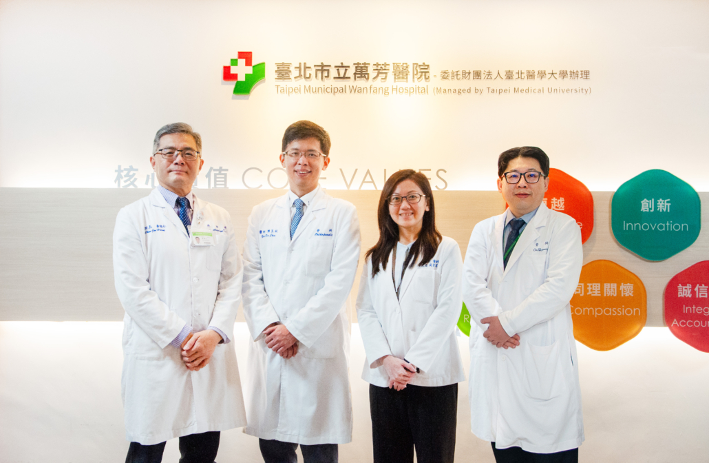 https://oge.tmu.edu.tw/wp-content/uploads/2024/03/骨科新創獎團隊合成-scaled.jpg
979
1500
global.initiatives
/wp-content/uploads/2018/05/02-紅logo並置.png
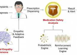
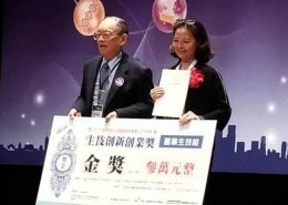
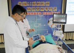
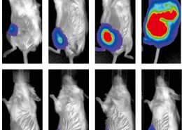
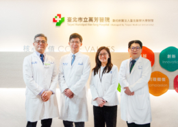
global.initiatives2024-04-03 09:00:252024-03-29 15:03:31TMU Healthcare System Develops Outstanding Innovative Medical Devices, Advancing AI Healthcare
https://oge.tmu.edu.tw/wp-content/uploads/2024/03/骨科新創獎團隊合成-scaled.jpg
979
1500
global.initiatives
/wp-content/uploads/2018/05/02-紅logo並置.png
global.initiatives2024-04-03 09:00:252024-03-29 15:03:31TMU Healthcare System Develops Outstanding Innovative Medical Devices, Advancing AI Healthcare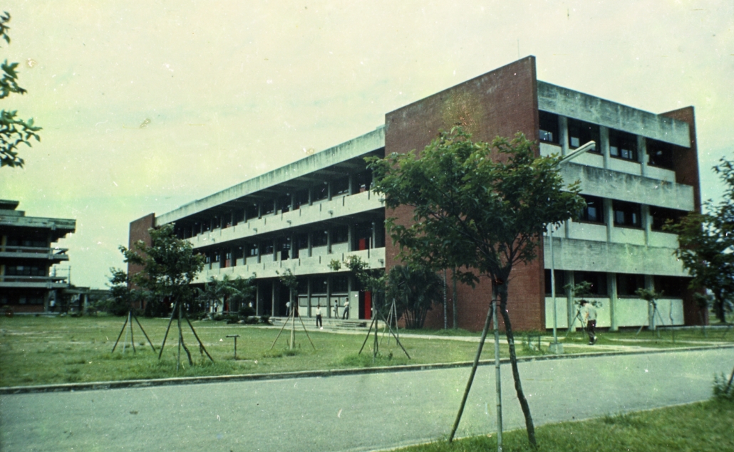 https://oge.tmu.edu.tw/wp-content/uploads/2024/03/feature-imageB074012-scaled.jpg
924
1500
global.initiatives
/wp-content/uploads/2018/05/02-紅logo並置.png
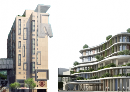
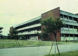
global.initiatives2024-04-02 09:00:322024-03-29 15:02:47Preserving History and Building Sustainability: The Renovation of TMU Teaching Building
https://oge.tmu.edu.tw/wp-content/uploads/2024/03/feature-imageB074012-scaled.jpg
924
1500
global.initiatives
/wp-content/uploads/2018/05/02-紅logo並置.png
global.initiatives2024-04-02 09:00:322024-03-29 15:02:47Preserving History and Building Sustainability: The Renovation of TMU Teaching Building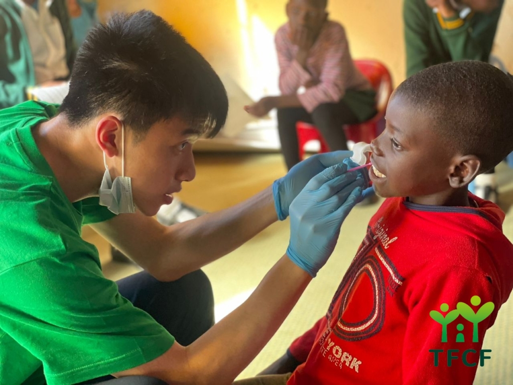 https://oge.tmu.edu.tw/wp-content/uploads/2024/03/史瓦新聞.jpg
960
1280
global.initiatives
/wp-content/uploads/2018/05/02-紅logo並置.png
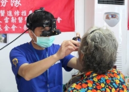
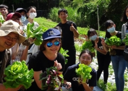
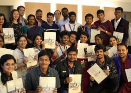
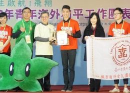
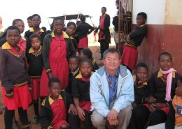
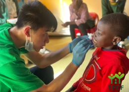
global.initiatives2024-03-28 09:00:182024-03-29 15:04:04TMU Students’ Overseas Volunteer Efforts Recognized by Taiwan’s Youth Development Administration
https://oge.tmu.edu.tw/wp-content/uploads/2024/03/史瓦新聞.jpg
960
1280
global.initiatives
/wp-content/uploads/2018/05/02-紅logo並置.png
global.initiatives2024-03-28 09:00:182024-03-29 15:04:04TMU Students’ Overseas Volunteer Efforts Recognized by Taiwan’s Youth Development Administration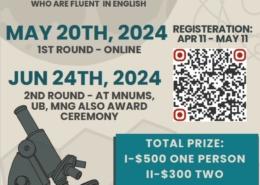


-260x185.jpg)


與連江縣衛生福利局陳美金局長簽署醫療合作備忘錄-260x185.jpg)


期許永續發展成為醫療產業新契機。-260x185.jpg)