TMU pioneers world’s largest virtual reality anatomy class
Source: Taipei Medical University
Published on 2019-05-03
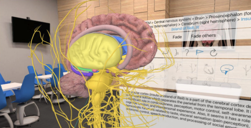
In collaboration with HTC, Taipei Medical University established the world’s largest virtual reality (VR) anatomy classroom in late 2018. The 3D Organon VR system gives headgear-wearing students a comprehensive view of structures in the human body.
The university is working with the health and medical division of smart phone and VR manufacturer HTC (High Tech Computer Corporation). The resulting world’s largest VR anatomy class is furnished with 10 sets of VIVE Pro (awarded 2018 VR headgear of the year) and 3D Organon VR anatomy software. This enables individual study as well as cooperative use of the same VR environment, and allows students to visualize lectures on anatomical structures in depth to better understand how bodies function
HTC
Anatomy and Cell Biology Director Hung-Ming Chang (張宏名) of the College of Medicine says adding VR to anatomy courses overcomes previous limitations such as unobservable structures or awkward angles. As students can better learn how anatomical structures are interlinked in three-dimensional spaces, this should increase instructional effectiveness.

Director Hung-Ming Chang
Director Chang calls the 3D Organon VR Anatomy software students’ best way to gain in-depth anatomical understanding. While cadaver dissection provides irreplaceable realism, the VR headset allows repeated examinations of various body parts unchanged by incisions and other exploratory changes that are part of cadaver study. Because the immersive 360-degree view shows tissues, bones, muscles, blood vessels, nerves and organs, students are better prepared for future clinical work.

Jerry Cheng (鄭志偉), HTC DeepQ Sr. Director, speaks and demonstrates VR headgear VIVE Pro
Edward Y. Chang (張智威), President of HTC DeepQ, says VR technology’s three-dimensional visualization is a new teaching method that accelerates learning. Use of VR in medical education and clinical applications will help more students, teachers, clinical medical staff and patients in the future.
Did you know? |
| Anatomy is the foundation of medicine, and learning through dissection provides essential training for medical students before clinical work. In the past, teaching materials for anatomy were primarily two-dimensional, such as textbooks, computer tablets and digital dissection tables. However, two-dimensional images require students to imagine how anatomical structures relate in three dimensions. |
Cadaver donations are limited and cannot be used repeatedly, limiting the time students have to work on a cadaver and learn dissection. The new 3D tools do not present these limitations.
The 3D Organon VR Anatomy system’s immersive learning environment increases student participation with different instructional techniques. It can support up to 300 individuals online at the same time, and can disassemble and rotate over 4000 anatomical structures in the VR environment. Besides stationary VR human body part models, the system also provides dynamic dissection models realistically presenting the extension and contraction of muscles and the beating of the heart. Even heart valve motion can be examined, giving students a perspective impossible to gain with a cadaver.
TMU anatomy classes all have VR equipment so students can study before, during and after dissection. They can move, disassemble or go through human body structures to note organs’ relative positions in combination with lab classes.
Future 3D Organon VR Anatomy improvements will complement TMU’s development of more VR courses that can be applied to previews, in-class use and reviews to encourage active learning. The system will be further expanded to in- service and continuing education, the “smart medicine” EMBA and medical camps for pre-college students.
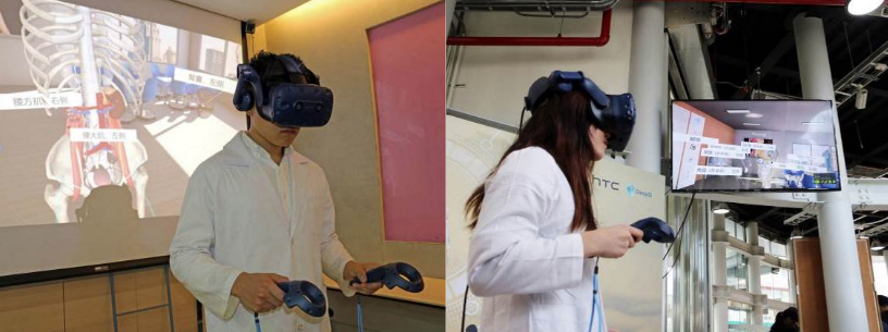
Students use VR equipment to study anatomy
For more relevant coverage, please click on the respective links as follows.
1. Forbes
2. QS WOWNews
 https://oge.tmu.edu.tw/wp-content/uploads/2024/04/olympiad-poster-2.jpg
909
640
gps
/wp-content/uploads/2018/05/02-紅logo並置.png
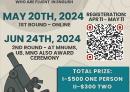
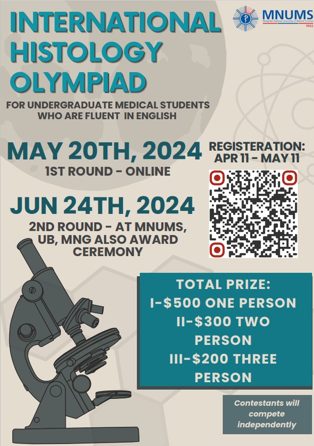
gps2024-04-19 16:36:372024-04-19 16:38:14International Histology Olympiad at the Mongolian National University of Medical Sciences (MNUMS)
https://oge.tmu.edu.tw/wp-content/uploads/2024/04/olympiad-poster-2.jpg
909
640
gps
/wp-content/uploads/2018/05/02-紅logo並置.png
gps2024-04-19 16:36:372024-04-19 16:38:14International Histology Olympiad at the Mongolian National University of Medical Sciences (MNUMS) https://oge.tmu.edu.tw/wp-content/uploads/2023/07/20230321台北醫學大學校園形象攝影-1-scaled.jpg
1000
1500
global.initiatives
/wp-content/uploads/2018/05/02-紅logo並置.png
global.initiatives2024-04-19 16:08:552024-04-19 16:59:58[Open Application] MOFA Taiwan Scholarship
https://oge.tmu.edu.tw/wp-content/uploads/2023/07/20230321台北醫學大學校園形象攝影-1-scaled.jpg
1000
1500
global.initiatives
/wp-content/uploads/2018/05/02-紅logo並置.png
global.initiatives2024-04-19 16:08:552024-04-19 16:59:58[Open Application] MOFA Taiwan Scholarship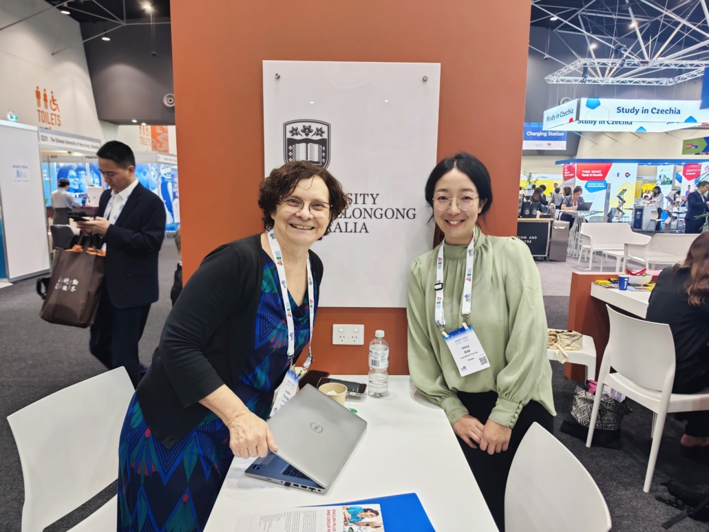 https://oge.tmu.edu.tw/wp-content/uploads/2024/04/1712888393174-scaled.jpg
1125
1500
gps
/wp-content/uploads/2018/05/02-紅logo並置.png
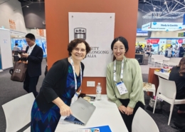
gps2024-04-15 16:31:512024-04-16 10:48:20Outward visit_2024 Asia Pacific Association of International Education (APAIE) conference and exhibition
https://oge.tmu.edu.tw/wp-content/uploads/2024/04/1712888393174-scaled.jpg
1125
1500
gps
/wp-content/uploads/2018/05/02-紅logo並置.png
gps2024-04-15 16:31:512024-04-16 10:48:20Outward visit_2024 Asia Pacific Association of International Education (APAIE) conference and exhibition https://oge.tmu.edu.tw/wp-content/uploads/2024/04/for-OGEW.jpg
788
940
global.initiatives
/wp-content/uploads/2018/05/02-紅logo並置.png
global.initiatives2024-04-12 12:05:012024-04-17 18:23:122024 TMU European Culture Festival
https://oge.tmu.edu.tw/wp-content/uploads/2024/04/for-OGEW.jpg
788
940
global.initiatives
/wp-content/uploads/2018/05/02-紅logo並置.png
global.initiatives2024-04-12 12:05:012024-04-17 18:23:122024 TMU European Culture Festival https://oge.tmu.edu.tw/wp-content/uploads/2024/04/shutterstock_1629801502.jpg
667
1000
global.initiatives
/wp-content/uploads/2018/05/02-紅logo並置.png
global.initiatives2024-04-10 17:42:292024-04-10 17:42:293 PhD programmes to get excited about
https://oge.tmu.edu.tw/wp-content/uploads/2024/04/shutterstock_1629801502.jpg
667
1000
global.initiatives
/wp-content/uploads/2018/05/02-紅logo並置.png
global.initiatives2024-04-10 17:42:292024-04-10 17:42:293 PhD programmes to get excited about https://oge.tmu.edu.tw/wp-content/uploads/2024/04/airmeet_da312f7f-4dfc-4912-8881-d0e302991d63.jpg
810
1440
gps
/wp-content/uploads/2018/05/02-紅logo並置.png
gps2024-04-09 15:00:432024-04-09 15:10:46Career Networking Event – working in Hungary
https://oge.tmu.edu.tw/wp-content/uploads/2024/04/airmeet_da312f7f-4dfc-4912-8881-d0e302991d63.jpg
810
1440
gps
/wp-content/uploads/2018/05/02-紅logo並置.png
gps2024-04-09 15:00:432024-04-09 15:10:46Career Networking Event – working in Hungary https://oge.tmu.edu.tw/wp-content/uploads/2024/03/骨科新創獎團隊合成-scaled.jpg
979
1500
global.initiatives
/wp-content/uploads/2018/05/02-紅logo並置.png
global.initiatives2024-04-03 09:00:252024-03-29 15:03:31TMU Healthcare System Develops Outstanding Innovative Medical Devices, Advancing AI Healthcare
https://oge.tmu.edu.tw/wp-content/uploads/2024/03/骨科新創獎團隊合成-scaled.jpg
979
1500
global.initiatives
/wp-content/uploads/2018/05/02-紅logo並置.png
global.initiatives2024-04-03 09:00:252024-03-29 15:03:31TMU Healthcare System Develops Outstanding Innovative Medical Devices, Advancing AI Healthcare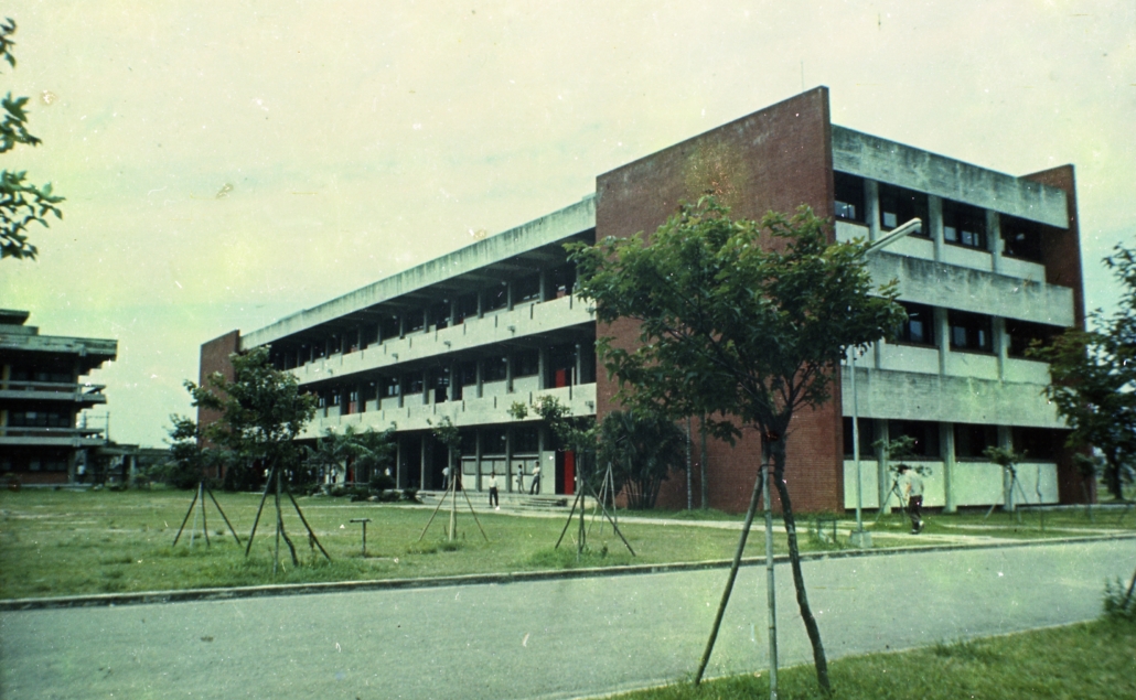 https://oge.tmu.edu.tw/wp-content/uploads/2024/03/feature-imageB074012-scaled.jpg
924
1500
global.initiatives
/wp-content/uploads/2018/05/02-紅logo並置.png
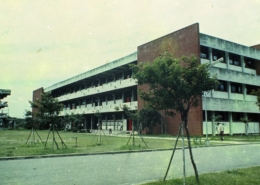
global.initiatives2024-04-02 09:00:322024-03-29 15:02:47Preserving History and Building Sustainability: The Renovation of TMU Teaching Building
https://oge.tmu.edu.tw/wp-content/uploads/2024/03/feature-imageB074012-scaled.jpg
924
1500
global.initiatives
/wp-content/uploads/2018/05/02-紅logo並置.png
global.initiatives2024-04-02 09:00:322024-03-29 15:02:47Preserving History and Building Sustainability: The Renovation of TMU Teaching Building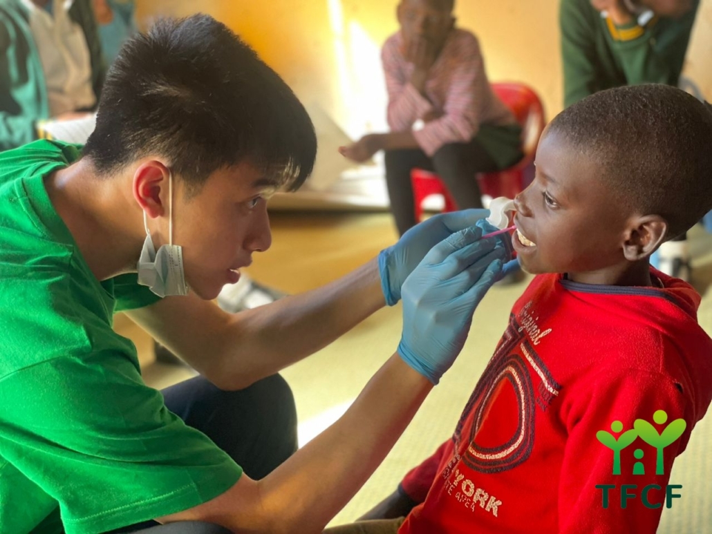 https://oge.tmu.edu.tw/wp-content/uploads/2024/03/史瓦新聞.jpg
960
1280
global.initiatives
/wp-content/uploads/2018/05/02-紅logo並置.png
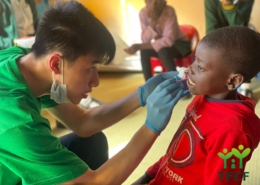
global.initiatives2024-03-28 09:00:182024-03-29 15:04:04TMU Students’ Overseas Volunteer Efforts Recognized by Taiwan’s Youth Development Administration
https://oge.tmu.edu.tw/wp-content/uploads/2024/03/史瓦新聞.jpg
960
1280
global.initiatives
/wp-content/uploads/2018/05/02-紅logo並置.png
global.initiatives2024-03-28 09:00:182024-03-29 15:04:04TMU Students’ Overseas Volunteer Efforts Recognized by Taiwan’s Youth Development Administration